Contamination in foot-and-mouth disease vaccine production: sources, impacts, and control strategies — A smart review
DOI:
https://doi.org/10.14295/bjs.v6i2.936Keywords:
foot-and-mouth disease, FMD vaccine, microbial contamination, vaccine manufacturing, biosafetyAbstract
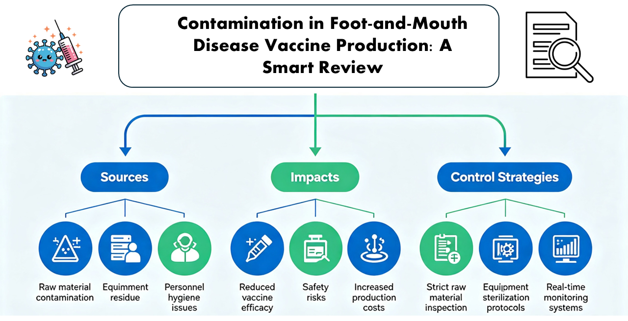
Foot-and-Mouth Disease (FMD) is a viral disease of cloven-hoofed animals that continues to pose serious threats to livestock health and the global economy. Inactivated vaccines continue to be the mainstay of an effective FMD control program, and much of the vaccine production relies on cell culture systems. Although improvements have been made in technology, contamination of FMD virus culture has remained a major limitation for the safety, potency, and reproducibility of vaccines. This review is a summary of current knowledge about types and sources of contamination observed in FMD vaccine production, such as bacterial, fungal, mycoplasma, and adventitious viral contamination. Aseptic handling of the product, sterilizations of equipment and reagents, reagent quality, and the manufacturing environment are highlighted, as these are common sources of contamination. The review will also address the effects contamination has on viral yield, antigen stability, and downstream vaccine quality. Moreover, the current detection, preventive, and quality control procedures at each phase of vaccine manufacture are critically reviewed. This review will focus on the existing issues and limitations in contaminant control, highlighting the areas that need to be addressed for enhanced consistency and biosafety of vaccines. We need to reinforce the overall effectiveness and reliability of FMD vaccination programs by improving contamination management.
References
Baaity, Z., Breunig, S., Önder, K., & Somogyvári, F. (2019). Direct qPCR is a sensitive approach to detect Mycoplasma contamination in U937 cell cultures. BMC Research Notes, 12, 720. https://doi.org/10.1186/s13104-019-4763-5
Barteling, S. J. (2002). Development and performance of inactivated vaccines against foot and mouth disease. Revue Scientifique et Technique-Office International des Épizooties, 21(3), 577-588.
Colenutt, C., Brown, E., Nelson, N., Paton, D. J., Eblé, P., Dekker, A., Gonzales, J. L., & Gubbins, S. (2020). Quantifying the transmission of foot-and-mouth disease virus in cattle via a contaminated environment. mBio, 11(4). https://doi.org/10.1128/mbio.00381-20
Corral-Vázquez, C., Aguilar-Quesada, R., Catalina, P., Lucena-Aguillar, G., Ligero, G., Miranda, B., & Carrillo-Ávila, J. A. (2017). Cell lines authentication and mycoplasma detection as minimum quality control of cell lines in biobanking. Cell and Tissue Banking, 18, 271-280. https://doi.org/10.1007/s10561-017-9617-6
Drexler, H. G., & Uphoff, C. C. (2002). Mycoplasma contamination of cell cultures: Incidence, sources, effects, detection, elimination, prevention. Cytotechnology, 39, 75-90. https://doi.org/10.1023/A:1022913015916
Feng, N., Huang, X., & Jia, Y. (2019). Mycoplasma contamination affects cell characteristics and decreases the sensitivity of BV2 microglia to LPS stimulation. Cytotechnology, 71, 623-634. https://doi.org/10.1007/s10616-019-00311-8
Gaboiphiwe, K., Kabelo, T. I., Mosholombe, T., Hyera, J., Fana, E. M., Masisi, K., & Lebani, K. (2025). A review of the utility of established cell lines for isolation and propagation of the southern african territories serotypes of foot-and-mouth disease virus. Viruses, 17(1), 39. https://doi.org/10.3390/v17010039
Gul, M., Kashif, M., Mihammad, S., Azizi, S., & Sun, H. (2025). Various methods of synthesis and applications of gold-based nanomaterials: a detailed review. Crystal Growth & Design, 25(7), 2227-2266. https://doi.org/10.1021/acs.cgd.4c01687
Hoff, F. W., Hu, C. W., Qutub, A. A., Qiu, Y., Graver, E., Hoang, G., Chauhan, M., de Bont, E. S. J. M., & Kornblau, S. M. (2018). Mycoplasma contamination of leukemic cell lines alters protein expression determined by reverse phase protein arrays. Cytotechnology, 70, 1529-1535. https://doi.org/10.1007/s10616-018-0244-2
Ishikawa, Y., Kozakai, T., Morita, H., Saida, K., Oka, S., & Masuo, Y. (2006). Rapid detection of mycoplasma contamination in cell cultures using SYBR Green-based real-time polymerase chain reaction. In Vitro Cellular & Developmental Biology – Animal, 42, 63-69. https://doi.org/10.1290/0505035.1
Kashif, M., Sharif, T., Khan, S., Shahzad, N., Amir, J., Gul, M., Shahid, K., Qarni, A., Shah, A., & Maninise, N. (2026). Synthesis strategies and multifaceted applications of molybdenum‐based nanomaterials: A Comprehensive review. Scientifica, 2026(1), 1764400. https://doi.org/10.1155/sci5/1764400
Kazemiha, V. M., Bonakdar, S., Amanzadeh, A., Azari, S., Memarnejadian, A., Shahbazi, S., Shokrgozar, M. A., & Mahdian, R. (2016). Real-time PCR assay is superior to other methods for the detection of mycoplasma contamination in the cell lines of the National Cell Bank of Iran. Cytotechnology, 68, 1063-1080. https://doi.org/10.1007/s10616-015-9862-0
Kazemiha, V. M., Shokrgozar, M. A., Arabestani, M. R., Moghadam, M. S., Azari, S., Maleki, S., & Amanzadeh, A. (2009). PCR-based detection and eradication of mycoplasmal infections from various mammalian cell lines: a local experience. Cytotechnology, 61(3), 117-124. https://doi.org/10.1007/s10616-010-9252-6
Khan, S., Hussain, Z., Bibi, S., Kashif, M., Zubair, M., & Azizi, S. (2026). Bias-free catalytic photoelectrochemical degradation of Congo red using a boron–iron oxide coated electrode: A green and sustainable. Catalysis Surveys from Asia, 30(1), 64-76. https://doi.org/10.1007/s10563-025-09465-2
Lawson-Ferreira, R., Santiago, M. A., Chometon, T. Q., Costa, V. A., Silva, S. A., Bertho, A. L., & Filipis, I. (2021). Flow-cytometric method for viability analysis of Mycoplasma gallisepticum and other cell-culture-contaminant Mollicutes. Current Microbiology, 78, 67-77. https://doi.org/10.1007/s00284-020-02255-1
Leonovich, O. A. (2024). Mycoplasma: Properties, detection, and decontamination methods of cell cultures and viral strains (Review). Applied Biochemistry and Microbiology, 60, 785-793. https://doi.org/10.1134/S0003683824604943
Olarerin-George, A. O., & Hogenesch, J. B. (2015). Assessing the prevalence of mycoplasma contamination in cell culture via a survey of NCBI's RNA-seq archive. Nucleic Acids Research, 43(5), 2535-2542. https://doi.org/10.1093/nar/gkv136
Radlett, P. J. (2005). The use of BHK suspension cells for the production of foot and mouth disease vaccines. Vertrebrate Cell Culture I, 129-146. https://doi.org/10.1007/BFb0000677
Shurson, G.C., P.E. Urriola, & Schroeder, D. C. (2023). Biosecurity and mitigation strategies to control swine viruses in feed ingredients and complete feeds. Animals,13(14), 2375. https://doi.org/10.3390/ani13142375
Soheily Z, Soleimani M, & Majidzadeh-Ardebili K. (2019). Detection of Mycoplasma contamination of cell culture by a loop-mediated isothermal amplification method. Cell Journal, 21(1), 43-48. https://doi.org/10.22074/cellj.2019.5624.
U.S. Food and Drug Administration (FDA). (2010). Characterization and Qualification of Cell Substrates and Other Biological Materials Used in the Production of Viral Vaccines for Infectious Disease Indications: Guidance for Industry. Silver Spring, MD: FDA.
Wehbe, K., Vezzalini, M., & Cinque, G. (2018). Detection of mycoplasma in contaminated mammalian cell culture using FTIR microspectroscopy. Analytical and Bioanalytical Chemistry, 410, 3003-3016. https://doi.org/10.1007/s00216-018-0987-9
Wirth, M., Berthold, E., Grashoff, M., Pfützner, H., Schubert, U., & Hauser, H. (1994). Detection of mycoplasma contaminations by the polymerase chain reaction. Cytotechnology, 16, 67-77. https://doi.org/10.1007/BF00754609
World Organisation for Animal Health (WOAH). (2022). Foot and mouth disease (infection with foot and mouth disease virus). Chapter 3.1.8. In Manual of Diagnostic Tests and Vaccines for Terrestrial Animals. World Organisation for Animal Health, Paris, France.
World Organisation for Animal Health (WOAH). (2024). Tests for sterility and freedom from contamination of biological materials intended for veterinary use. Chapter 1.1.9. In Manual of Diagnostic Tests and Vaccines for Terrestrial Animals. World Organisation for Animal Health, Paris, France.
World Health Organization (WHO). (2016). WHO good manufacturing practices for biological products. Annex 2. WHO Technical Report Series No. 999. World Health Organization, Geneva, Switzerland.
World Organisation for Animal Health (WOAH). (2022). Principles of veterinary vaccine production. Chapter 1.1.8. In Manual of Diagnostic Tests and Vaccines for Terrestrial Animals. World Organisation for Animal Health, Paris, France. This chapter provides additional guidance on starting materials, contamination prevention, and vaccine manufacturing controls.
World Organisation for Animal Health (WOAH). (2022). Foot and mouth disease (infection with foot and mouth disease virus). Chapter 3.1.8. In: Manual of Diagnostic Tests and Vaccines for Terrestrial Animals. Paris: WOAH.
World Health Organization (WHO). (2013). Recommendations for the evaluation of animal cell cultures as substrates for the manufacture of biological medicinal products and for the characterization of cell banks. WHO Technical Report Series No. 978, Annex 3. Geneva: WHO.
Yu, T., Wang, Y., Zhang, H., Johnson, C. H., Jiang, Y., Li, X., Wu, Z., Liu, T., Huang, M., & Bi, H. (2016). Metabolomics reveals mycoplasma contamination interferes with the metabolism of PANC-1 cells. Analytical and Bioanalytical Chemistry, 408, 4267-4273. https://doi.org/10.1007/s00216-016-9525-9
Downloads
Published
How to Cite
Issue
Section
License
Copyright (c) 2027 Muhammad Atif, Muhammad Shahid, Ayessha Gohar, Zainab Liaqat, Muhammad Usman Ali Malik

This work is licensed under a Creative Commons Attribution 4.0 International License.
Authors who publish with this journal agree to the following terms:
1) Authors retain copyright and grant the journal right of first publication with the work simultaneously licensed under a Creative Commons Attribution License that allows others to share the work with an acknowledgement of the work's authorship and initial publication in this journal.
2) Authors are able to enter into separate, additional contractual arrangements for the non-exclusive distribution of the journal's published version of the work (e.g., post it to an institutional repository or publish it in a book), with an acknowledgement of its initial publication in this journal.
3) Authors are permitted and encouraged to post their work online (e.g., in institutional repositories or on their website) prior to and during the submission process, as it can lead to productive exchanges, as well as earlier and greater citation of published work.