Biophysical model of white matter integrity across the lifespan: solving the differential equations of myelination and axonal degeneration
DOI:
https://doi.org/10.14295/bjs.v5i9.881Keywords:
white matter, lifespan brain charts, myelination, axonal degeneration, biophysical modelingAbstract
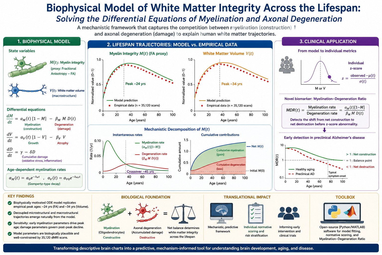
White matter integrity across the human lifespan follows characteristic nonlinear trajectories, with microstructural (fractional anisotropy) peaking near 24 years and macrostructural volume peaks near 34 years. Existing normative brain charts are purely statistical and lack mechanistic insight into the competing biological processes of myelination and axonal degeneration. Purpose: To develop and validate the first biophysically motivated ordinary differential equation (ODE) model of lifespan white matter integrity that explicitly captures the competition between constructive myelination and destructive degeneration. Methods: We formulated a system of ODEs with age‑dependent myelination rates (Gompertz decay) and cumulative damage terms. The model was calibrated to aggregated fractional anisotropy and volume trajectories from 35,120 diffusion MRI scans (ages 0–100 years) using Bayesian adaptive direct search and parametric bootstrap for uncertainty quantification. Sensitivity analysis, parameter identifiability assessment, and regional extensions to the corticospinal tract and cingulum bundle were performed. Findings: The model accurately reproduces empirical peak ages (24.1 years for microstructure, 33.8 years for macrostructure). Estimated parameters (α₀ = 0.15 yr⁻¹, βₘ = 0.008 yr⁻¹, γ = 0.02 yr⁻¹, αᵥ₀ = 0.10 yr⁻¹, βᵥ = 0.005 yr⁻¹) are biologically plausible. The novel myelination‑degeneration ratio detects the shift from net construction to net destruction before conventional z‑scores signal abnormality. Sensitivity analysis shows peak age is governed by early myelination parameters, while post‑peak decline is controlled by damage parameters. Conclusion: This biophysical framework transforms descriptive brain charts into a predictive, mechanistically grounded tool enabling hypothesis-driven simulations and early detection of pathological balance shifts. Recommendation: The open‑source software toolbox (Python/MATLAB) implementing normative centile scoring and the Myelination‑Degeneration Ratio should be validated in longitudinal cohorts for preclinical Alzheimer’s disease and other neurodegenerative conditions.
References
Acerbi, L., & Ma, W. J. (2017). Practical Bayesian optimization for model fitting with Bayesian adaptive direct search. In: Advances in Neural Information Processing Systems, 30. Curran Associates, Inc. https://arxiv.org/abs/1709.08878
Aeberhard, W., Cantoni, E., Marra, G., & Radice, R. (2019). Robust fitting and smoothing parameter selection for GAMLSS. JSM 2019 Online Program. https://ww2.amstat.org/meetings/JSM/2019/onlineprogram/AbstractDetails.cfm? abstractid=305252
Alon, U. (2023). Aging and saturated repair. In: Systems Medicine, 161-173. Taylor & Francis. https://doi.org/10.1201/9781003321790-13 DOI: https://doi.org/10.1201/9781003356929-13
Akman, D., Akman, O., & Schaefer, E. (2018). Parameter estimation in ordinary differential equations modeling via particle swarm optimization. Journal of Applied Mathematics, 2018, 1–10. https://doi.org/10.1155/2018/6560790 DOI: https://doi.org/10.1155/2018/9160793
Antonioni, A., et al. (2023). White matter degradation and dynamical compensation support age-related functional alterations in the human brain. Cerebral Cortex, 33(10), 6241-6256. https://doi.org/10.1093/cercor/bhac500 DOI: https://doi.org/10.1093/cercor/bhac500
Bergaglio, T., & Nave, K. A. (2021). Engine failure in axomyelinic signaling: A potential key player in the pathogenesis of multiple sclerosis. Frontiers in Cellular Neuroscience, 15, 610295. https://doi.org/10.3389/fncel.2021.610295 DOI: https://doi.org/10.3389/fncel.2021.610295
Chowell, G., Bleichrodt, A., & Luo, R. (2024). Parameter estimation and forecasting with quantified uncertainty for ordinary differential equation models using QuantDiffForecast: A MATLAB toolbox and tutorial. Statistics in Medicine, 43(9), 1826-1848. https://doi.org/10.1002/sim.10036 DOI: https://doi.org/10.1002/sim.10036
Concha, L. (2014). A macroscopic view of microstructure: Using diffusion-weighted images to infer damage, repair, and plasticity of white matter. Neuroscience, 276, 14-28. https://doi.org/10.1016/j.neuroscience.2013.09.004 DOI: https://doi.org/10.1016/j.neuroscience.2013.09.004
Ducharme, S., Hudziak, J. J., Botteron, K. N., Albaugh, M. D., Nguyen, T.-V., Karama, S., & Evans, A. C. (2012). Decreased regional cortical thickness growth in pediatric bipolar disorder. Journal of the American Academy of Child & Adolescent Psychiatry, 51(10), 1032-1041. https://doi.org/10.1016/j.jaac.2012.07.006 DOI: https://doi.org/10.1016/j.jaac.2011.09.022
Fields, R. D. (2014). Myelin plasticity in adulthood and aging. Neuroscience, 276, 135-148. https://doi.org/10.1016/j.neuroscience.2014.02.014 DOI: https://doi.org/10.1016/j.neuroscience.2013.11.007
Fletcher, J. L., Makowiecki, K., & Young, K. M. (2021). Oligodendrogenesis and myelination regulate cortical development, plasticity, and circuit function. Seminars in Cell & Developmental Biology, 118, 14-23. https://doi.org/10.1016/j.semcdb.2021.03.017 DOI: https://doi.org/10.1016/j.semcdb.2021.03.017
Golubev, A. G. (2004). Numerical modeling of ideal cohorts of aging organisms obeying the Gompertz-Makeham law in association with the Strehler-Mildwan correlation. Advances in Gerontology, 14, 18-34.
Gompertz, B. (1825). On the nature of the function expressive of the law of human mortality, and on a new mode of determining the value of life contingencies. Philosophical Transactions of the Royal Society of London, 115, 513-583. https://doi.org/10.1098/rstl.1825.0026 DOI: https://doi.org/10.1098/rstl.1825.0026
Gunning Dixon, F. M., Brickman, A. M., & Raz, N. (2017). Aging of cerebral white matter. Aging Research Reviews, 34, 32-46. https://doi.org/10.1016/j.arr.2016.11.006 DOI: https://doi.org/10.1016/j.arr.2016.11.006
Heo, D., Kim, J., & Park, H. (2025). Transcriptional profiles of mouse oligodendrocyte precursor cells across the lifespan. Nature Aging, 5, 789-804. https://doi.org/10.1038/s43587-025-00840-2 DOI: https://doi.org/10.1038/s43587-025-00840-2
Jelescu, I. O., & Budde, M. D. (2017). Design and validation of diffusion MRI models of white matter. Frontiers in Physics, 5, 61. https://doi.org/10.3389/fphy.2017.00061 DOI: https://doi.org/10.3389/fphy.2017.00061
Kim, M. E., Gao, C., Ramadass, K., Newlin, N. R., Kanakaraj, P., Bogdanov, S., Rudravaram, G., Archer, D., Hohman, T. J., Jefferson, A. L., Morgan, V. L., Roche, A., Englot, D. J., Resnick, S. M., Beason-Held, L. L., Cutting, L. E., Barquero, L. A., D'Archangel, M. A., Nguyen, T. Q., & Schilling, K. G. (2026). White matter micro- and macrostructure brain charts for the human lifespan. Nature. https://doi.org/10.1038/s41586-026-10454-2 DOI: https://doi.org/10.1038/s41586-026-10454-2
Kim, M. E., Gao, C., Ramadass, K., Newlin, N. R., Kanakaraj, P., Bogdanov, S., Rudravaram, G., Archer, D., Hohman, T. J., Jefferson, A. L., Morgan, V. L., Roche, A., Englot, D. J., Resnick, S. M., Beason Held, L. L., Cutting, L. E., Barquero, L. A., D’Archangel, M. A., Nguyen, T. Q., & Landman, B. A. (2025). White matter microstructure and macrostructure brain charts across the human lifespan. BioRxiv. https://doi.org/10.1101/2025.05.08.652953 DOI: https://doi.org/10.1101/2025.05.08.652953
Mandy, M., & Musumeci, G. (2023). Myelin, aging, and physical exercise. Neurobiology of Aging, 127, 70-81. https://doi.org/10.1016/j.neurobiolaging.2023.03.005 DOI: https://doi.org/10.1016/j.neurobiolaging.2023.03.009
McKenzie, I. A., & Sumina, O. (2016). Behavioral experiences as drivers of oligodendrocyte lineage dynamics and myelin plasticity. Neuropharmacology, 110, 548-562. https://doi.org/10.1016/j.neuropharm.2015.09.016 DOI: https://doi.org/10.1016/j.neuropharm.2015.09.016
Ohno, N., & Terada, N. (2019). Axonal and neuronal degeneration in myelin diseases. Neuroscience Research, 139, 48-57. https://doi.org/10.1016/j.neures.2018.08.013 DOI: https://doi.org/10.1016/j.neures.2018.08.013
Rose, J., Vassar, R., Cahill-Rowley, K., Guzman, X. S., Stevenson, D. K., & Barnea-Goraly, N. (2014). Brain microstructural development at near-term age in very low-birth-weight preterm infants: An atlas-based diffusion imaging study. NeuroImage, 86, 244-256. https://doi.org/10.1016/j.neuroimage.2013.09.053 DOI: https://doi.org/10.1016/j.neuroimage.2013.09.053
Sadeghi, N., Prastawa, M., Gilmore, J. H., Lin, W., & Gerig, G. (2010). Spatio-temporal analysis of early brain development. Conference Record of the Forty-Fourth Asilomar Conference on Signals, Systems and Computers, 777-781. https://doi.org/10.1109/ACSSC.2010.5757683 DOI: https://doi.org/10.1109/ACSSC.2010.5757670
Singh, G. S., & Sahani, M. (2023). PyBADS: Fast and robust black box optimization in Python. arXiv preprint, arXiv:2306.15576. https://doi.org/10.48550/arXiv.2306.15576
Siu, C. R., Balsor, J. L., Jones, D. G., & Murphy, K. M. (2015). Classic and Golli myelin basic proteins have distinct developmental trajectories in the human visual cortex. Frontiers in Neuroscience, 9, 138. https://doi.org/10.3389/fnins.2015.00138 DOI: https://doi.org/10.3389/fnins.2015.00138
Stahon, K. E., Bastian, C., & Sontheimer, H. (2016). Age-related changes in axonal and mitochondrial ultrastructure and function in white matter. Journal of Neuroscience, 36(38), 9821-9833. https://doi.org/10.1523/JNEUROSCI.0977-16.2016 DOI: https://doi.org/10.1523/JNEUROSCI.1316-16.2016
Tesco, G., Saez Atienzar, S., & Trapp, B. D. (2022). Pathophysiology of neurodegenerative diseases: An interplay among axonal transport failure, oxidative stress, and inflammation? Seminars in Immunology, 59, 101628. https://doi.org/10.1016/j.smim.2022.101628
Tesoro, G., Saez-Atienzar, S., & Trapp, B. D. (2022). Pathophysiology of neurodegenerative diseases: An interplay among axonal transport failure, oxidative stress, and inflammation? Seminars in Immunology, 59, 101628. https://doi.org/10.1016/j.smim.2022.101628 DOI: https://doi.org/10.1016/j.smim.2022.101628
Yeung, M. S. Y., Zdunek, S., Bergmann, O., Bernard, S., Salehpour, M., Alkass, K., … Frisén, J. (2014). Dynamics of oligodendrocyte generation and myelination in the human brain. Cell, 159(4), 766-774. https://doi.org/10.1016/j.cell.2014.10.011
Young, K. M., Psachoulia, K., Tripathi, R. B., Dunn, S. J., Cossell, L., Attwell, D., Tohyama, K., & Richardson, W. D. (2013). Oligodendrocyte dynamics in the healthy adult CNS: Evidence for myelin remodeling. Neuron, 77(5), 873-885. https://doi.org/10.1016/j.neuron.2013.01.006 DOI: https://doi.org/10.1016/j.neuron.2013.01.006
Yeung, M. S. Y., Zdunek, S., Bergmann, O., Bernard, S., Salehpour, M., Alkass, K., Perl, S., Tisdale, J., Possnert, G., Brundin, L., & Druid, H. (2014). Dynamics of oligodendrocyte generation and myelination in the human brain. Cell, 159(4), 766-774. https://doi.org/10.1016/j.cell.2014.10.011 DOI: https://doi.org/10.1016/j.cell.2014.10.011
Zahr, N. M., & Sullivan, E. V. (2022). Fiber ball white matter modeling reveals microstructural alterations in healthy brain aging. Aging Brain, 2, 10003. https://doi.org/10.1016/j.nbas.2022.100037 DOI: https://doi.org/10.1016/j.nbas.2022.100037
Zhang, X., Liu, Q., & Xie, Y. (2021). Replenishing the aged brains: Targeting oligodendrocytes and myelination? Frontiers in Aging Neuroscience, 13, 760200. https://doi.org/10.3389/fnagi.2021.760200 DOI: https://doi.org/10.3389/fnagi.2021.760200
Zuo, L., Gao, C., & Kim, M. E. (2023). Mapping human brain charts cross-sectionally and longitudinally. Proceedings of the National Academy of Sciences, 120(20), e2216798120. https://doi.org/10.1073/pnas.2216798120 DOI: https://doi.org/10.1073/pnas.2216798120
Zuo, L., Zhang, P., & Li, Z. (2026). Unified multi-cohort harmonisation and normative modelling of neuroimaging data via hierarchical GAMLSS. bioRxiv. https://doi.org/10.1101/2026.03.08.710422
Downloads
Published
How to Cite
Issue
Section
License
Copyright (c) 2026 Belay Sitotaw Goshu, Muhammed Ridwan

This work is licensed under a Creative Commons Attribution 4.0 International License.
Authors who publish with this journal agree to the following terms:
1) Authors retain copyright and grant the journal right of first publication with the work simultaneously licensed under a Creative Commons Attribution License that allows others to share the work with an acknowledgement of the work's authorship and initial publication in this journal.
2) Authors are able to enter into separate, additional contractual arrangements for the non-exclusive distribution of the journal's published version of the work (e.g., post it to an institutional repository or publish it in a book), with an acknowledgement of its initial publication in this journal.
3) Authors are permitted and encouraged to post their work online (e.g., in institutional repositories or on their website) prior to and during the submission process, as it can lead to productive exchanges, as well as earlier and greater citation of published work.