
Adapted silver impregnation protocol for the detection of Merkel cells in fish skin embedded in plastic historesins
DOI:
https://doi.org/10.14295/bjs.v5i1.813Keywords:
argyrophil silver impregnation, fish skin, histologic resin, light microscopy, Merkel cellAbstract
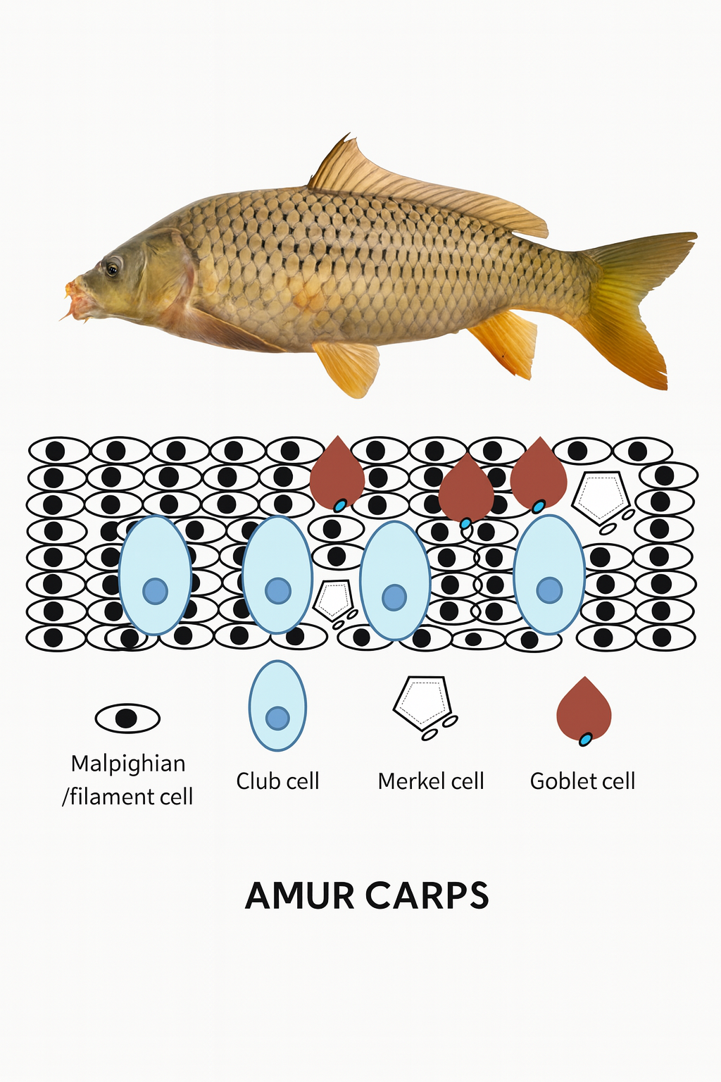
Merkel cells are detected using silver impregnation in skin samples embedded in paraplast. Here, we describe an adapted protocol for detecting Merkel cells in Amur carp (Cyprinus rubrofuscus) skin samples embedded in historesin. Incubated slides in Grimelius silver impregnation solution at 60 ºC for five hours resulted in positive Merkel cells scattered among skin cell layers composed of Malpighian, goblet, and club cells. Thus, Merkel cells can be explored in samples embedded in historesins using adapted protocols for paraplast ones.
References
AVMA. (2013). American Veterinary Medical Association. AVMA guidelines for the euthanasia of animals: 2013 edition. Schaumburg, IL: American Veterinary Medical Association.
Bogutskaya, N. G., Naseka, A. M., Shedko, S. V., Vasileva, E. D., & Chereshnev, I. A. (2008). The fishes of the Amur River: updated check-list and zoogeography. Ichthyological Exploration of Freshwaters, 4, 301-366
Day, R., & Salzet, M. (2002). The neuroendocrine phenotype, cellular plasticity, and the search for genetic switches: redefining the diffuse neuroendocrine system. Neuro Endocrinology Letters, 23, 447-451
DeLellis, R. A. (2001). The neuroendocrine system and its tumors. American Journal of Clinical Pathology, 115, S5-S16. https://doi.org/10.1309/7GR5-L7YW-3G78-LDJ6 DOI: https://doi.org/10.1309/7GR5-L7YW-3G78-LDJ6
Grimelius, L. (2004). Silver stains demonstrating neuroendocrine cells. Biotechnic & Histochemistry, 79, 37-44. https://doi.org/10.1080/10520290410001715264 DOI: https://doi.org/10.1080/10520290410001715264
Grimelius, L. (2008). Methods in neuroendocrine histopathology, a methodological overview. Upsala Journal of Medical Sciences, 113, 243-260. https://doi.org/10.3109/2000-1967-238 DOI: https://doi.org/10.3109/2000-1967-238
Halata, Z., Grim, M., & Bauman, K. I. (2003). Friedrich Sigmund Merkel and his “Merkel cell”, morphology, development, and physiology: review and new results. The Anatomical Record: Part A, Discoveries in Molecular, Cellular, and Evolutionary Biology, 271, 225-239. https://doi.org/10.1002/ar.a.10029 DOI: https://doi.org/10.1002/ar.a.10029
Iger, Y., Lock, R. A. C., Jenner, H. A., & Wendelaar Bonga, S. E. (1994a). Cellular responses in the skin of carp (Cyprinus carpio) exposed to copper. Aquatic Toxicology, 29, 49-64. https://doi.org/10.1016/0166-445X(94)90047-7 DOI: https://doi.org/10.1016/0166-445X(94)90047-7
Iger, Y., Lock, R. A. C., van der Meij, J. C. A., & Wendelaar Bonga, S. E. (1994b). Effects of Water-Borne Cadmium on the Skin of the Common Carp (Cyprinus carpio). Archives of Environmental Contamination and Toxicology, 26, 342-350. https://doi.org/10.1007/BF00203561 DOI: https://doi.org/10.1007/BF00203561
Iger, Y., Wendelaar Bonga, S. E. (1994). Cellular responses of the skin of carp (Cyprinus carpio) exposed to acidified water. Cell and Tissue Research, 275, 481-492. https://doi.org/10.1007/BF00318817 DOI: https://doi.org/10.1007/BF00318817
Kasumyan, A. O. (2011). Tactile reception and behavior of fish. Journal of Ichthyology, 51, 1035-1103. https://doi.org/10.1134/S003294521111004X DOI: https://doi.org/10.1134/S003294521111004X
Kotrschal, K., Whitear, M., & Finger, T. E. (1993). Spinal and facial innervation of the skin in the gadid fish Ciliata mustela (Teleostei). Journal of Comparative Neurology, 331, 407-417. https://doi.org/10.1002/cne.903310310 DOI: https://doi.org/10.1002/cne.903310310
Lane, E. B., & Whitear, M. (1977). On the occurrence of Merkel cells in the epidermis of teleost fishes. Cell and Tissue Research, 182:235-246. https://doi.org/10.1007/BF00220592 DOI: https://doi.org/10.1007/BF00220592
Lundqvist, M., Arnberg, H., Candell, J., Malmgren, M., Wilander, E., Grimelius, L., & Öberg, K. (1990). Silver stains for identification of neuroendocrine cells. A study of the chemical background. Histochemical Journal, 22, 615-623. https://doi.org/10.1007/BF01072943 DOI: https://doi.org/10.1007/BF01072943
Mazzoni, T. S., & Quagio-Grassiotto, I. (2020). In totum immunostaining: A histological analysis tool for small dimensions biological samples. International Journal of Biological and Medical Research, 11, 6938-6943
Mazzoni, T. S., & Quagio-Grassiotto, I. (2021). Presence of the matrix metalloproteinases during the migration of the primordial germ cells in zebrafish gonadal ridge. Cell and Tissue Research, 383, 707-722. https://doi.org/10.1007/s00441-020-03288-5 DOI: https://doi.org/10.1007/s00441-020-03288-5
Montuenga, L. M., Guembe, L., Burrell, A., Bodegas, M. E., Calvo, A., Sola, J. J., Sesma, P., & Villaro, A. C. (2003). The diffuse endocrine system: from embryogenesis to carcinogenesis. Progress in Histochemistry and Cytochemistry, 38, 155-272. https://doi.org/10.1016/S0079-6336(03)80004-9 DOI: https://doi.org/10.1016/S0079-6336(03)80004-9
Nurse, C. A., & Farraway, L. (1989). Characterization of Merkel cells and mechanosensory axons of the rat by styryl pyridinium dyes. Cell and Tissue Research, 255, 125-128. https://doi.org/10.1007/BF00229073 DOI: https://doi.org/10.1007/BF00229073
Rajan, S. T., & Malathi, N. (2014). Health hazards of xylene: a literature review. Journal of Clinical and Diagnostic Research, 8, 271-274. https://doi.org/10.7860/JCDR/2014/7544.4079 DOI: https://doi.org/10.7860/JCDR/2014/7544.4079
Rakers, S., Gebert, M., Uppalapati, S., Meyer, W., Maderson, P., Sell, A. F., Kruse, C., & Paus, R. (2010). ‘Fish matters’: the relevance of fish skin biology to investigative dermatology. Experimental Dermatology, 19, 313-324. https://doi.org/10.1111/j.1600-0625.2009.01059.x DOI: https://doi.org/10.1111/j.1600-0625.2009.01059.x
Ramírez, G. A., & de los Monteros, A. E. (2019). Study on the role of histochemical stains in identifying Merkel cells in dogs. The Anatomical Records, 302, 1458-1464. https://doi.org/10.1002/ar.24013 DOI: https://doi.org/10.1002/ar.24013
Shephard, K. L. (1994). Functions for fish mucus. Reviews in Fish Biology and Fisheries, 4, 401-429. https://doi.org/10.1007/BF00042888 DOI: https://doi.org/10.1007/BF00042888
Slominski, A. (2005). Neuroendocrine system of the skin. Dermatology, 211, 199-208. https://doi.org/10.1159/000087012 DOI: https://doi.org/10.1159/000087012
Tachibana, T., Ishizeki, K., Sakakura, Y., & Nawa, T. (1984). Ultrastructural evidence for a possible secretory function of Merkel cells in the barbels of a teleost fish, Cyprinus carpio. Cell and Tissue Research, 235, 695-697. https://doi.org/10.1007/BF00226971 DOI: https://doi.org/10.1007/BF00226971
Tachibana, T., & Nawa, T. (2002). Recent progress in studies on Merkel cell biology. Anatomical Science International, 77, 26-33. https://doi.org/10.1046/j.0022-7722.2002.00008.x DOI: https://doi.org/10.1046/j.0022-7722.2002.00008.x
Tachibana, T. (2005). The Merkel cell: recent findings and unresolved problems. Archives of Histology and Cytology, 58, 379-396. https://doi.org/10.1679/aohc.58.379 DOI: https://doi.org/10.1679/aohc.58.379
Weihe, E., Hartschuh, W., Schäfer, M. K., Romeo, H., & Elden, L. E. (1998). Cutaneous Merkel cells of the rat contain both dynorphin A and vesicular monoamine transporter type 1 (VMAT1) immunoreactivity. Canadian Journal of Physiology and Pharmacology, 76, 334-339. https://doi.org/10.1139/y98-032 DOI: https://doi.org/10.1139/y98-032
Whitear, M., & Lane, E. B. (1981). Fine structure of Merkel cells in lamprey. Cell and Tissue Research, 220, 139-151. https://doi.org/10.1007/BF00209973 DOI: https://doi.org/10.1007/BF00209973
Zaccone, G. (1986). Neuron-specific enolase and serotonin in the Merkel cells of conger-eel (Conger conger) epidermis: An immunohistochemical study. Histochemistry, 85, 29-34. https://doi.org/10.1007/BF00508650. DOI: https://doi.org/10.1007/BF00508650
Zaccone, G., Fasulo, S., & Ainis, L. (1994). Distribution cells in the determined histological patterns of the paraneuronal endocrine skin, gills and the airways of fishes as by immunohistochemical and methods. Histochemical Journal, 26, 609-629. https://doi.org/10.1007/BF00158286 DOI: https://doi.org/10.1007/BF00158286
Zhou, J., Wu, Q., Wang, Z., & Ye, Y. (2004). Molecular phylogeny of three subspecies of common carp Cyprinus carpio, based on sequence analysis of cytochrome b and control region of mtDNA. Journal of Zoological Systematics and Evolutionary Research, 42, 266-269. https://doi.org/10.1111/j.1439-0469.2004.00266.x DOI: https://doi.org/10.1111/j.1439-0469.2004.00266.x
Downloads
Published
How to Cite
Issue
Section
License
Copyright (c) 2026 André Luiz Veiga Conrado, José Roberto Machado Cunha da Silva

This work is licensed under a Creative Commons Attribution 4.0 International License.
Authors who publish with this journal agree to the following terms:
1) Authors retain copyright and grant the journal right of first publication with the work simultaneously licensed under a Creative Commons Attribution License that allows others to share the work with an acknowledgement of the work's authorship and initial publication in this journal.
2) Authors are able to enter into separate, additional contractual arrangements for the non-exclusive distribution of the journal's published version of the work (e.g., post it to an institutional repository or publish it in a book), with an acknowledgement of its initial publication in this journal.
3) Authors are permitted and encouraged to post their work online (e.g., in institutional repositories or on their website) prior to and during the submission process, as it can lead to productive exchanges, as well as earlier and greater citation of published work.